The new babies are here
I got a little Christmas money, and prepared to have some fun while waiting for hiking weather to return.
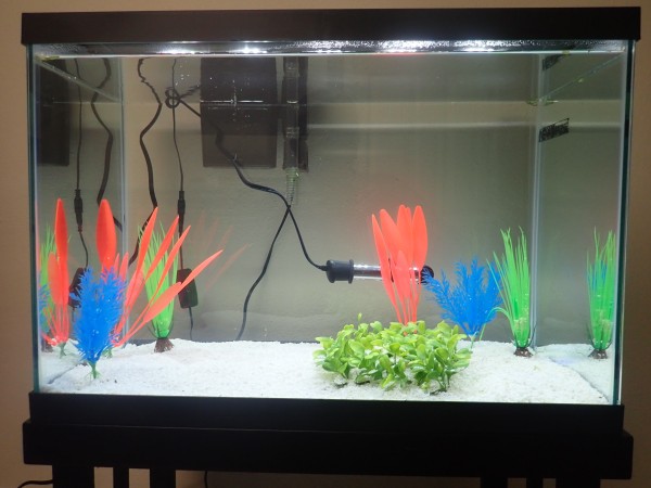
Twenty-gallon fish tank. I actually plan to use this as a quarantine tank, which I badly need, but which is not as much fun as a display tank. I consoled myself by promising myself a 10-gallon shrimp tank for my birthday, in three months.
I’m now cycling the quarantine tank with ammonium chloride. Since the tank will only occasionally have fish in it, I’ll have to keep the biological filter alive with an artificial ammonium source. Five days and no signs of cycling yet, but it happens quickly when it finally happens.
The other new baby:
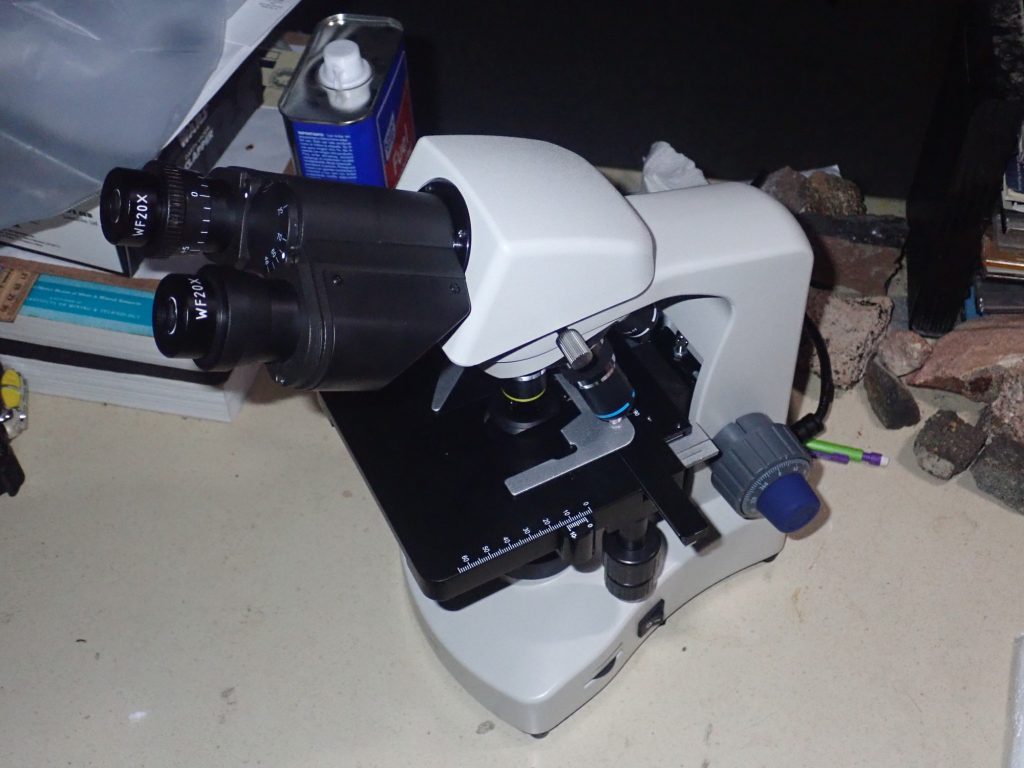
A new AmScope compound microscope with dark field capability.
I had a cheap microscope as a kid that had one objective already cracked when I got it, another with obvious astigmatism, and a highest-power objective that needed to be oil immersion but wasn’t. I had loads of fun with it anyway. It disappeared sometime in the last twenty years, I suspect when I moved to Los Alamos 17 years ago. I’m recapturing some childhood here.
I’m having a tremendous amount of fun with it. I pulled some gunk out of my fish tand and so far I’ve observed some Colpoda and other ciliated protozoans, some nematodes, some Vorticella, a colony of Stentor, and rotifers. Lots of rotifers. They seem to like attaching to half-dead plant matter. Their little wheels of cilia they filter feed with are gorgeous in the dark field. Also saw some Blepharisma, a new one to me, which my wife had to identify. (She’s the professional biologist.) Also some stomata on a live plant leaf.
And a blood smear. My own blood, of course. Not a problem getting a sample when you’re a diabetic. It took three tries to get a good stained smear. The first smeared okay, but I got overambitious with the stain; I don’t have any Wright’s stain or eosin, and tried counterstaining methylene blue with carbolic fuchsin, and they did not react well. Second smear was smeared badly. Third smeared okay, the methanol fixative worked, and I gave it a good ten minutes for the methylene blue to soak it and did not try to counterstain. So I would not be able to tell eosinophils from basophils from neutrophils. No matter. I could distinguish polymorphonuclear leukocytes (try saying that three times fast) from monocytes, and it was way cool. The nuclei showed up very clearly. I tried the oil immersion lens on it but that didn’t seem to accomplish much and the oil ruined the slide. Still.
I didn’t have any great excess of white cells — I had to hunt around for them — which is comforting. On the other hand, there seemed to be more monocytes than PMNs, when the ratio should be 2:1 in favor of PMNs. Maybe I’ve got mono. I’ve had a persistent headache since before Christmas (worse when I cough, bend over, or otherwise strain) but no other symptoms except some congestion and, today, a somewhat sore throat. I saw the PA at my doctor’s office Monday and she kind of scratched her head and said if it didn’t clear up in a couple of weeks, she’d order up some imaging. Would like to avoid that.
Saw some blue clumps that might have been clumps of platelets. This was blood straight from my finger, without anticoagulant, and platelets gonna plate.
Regardless. In addition to practicing medicine on myself without a license, I’ve just had loads of fun with the new scope. A stereo microscope would have been better for geology, but that’s what loupes are for, and I have looked at some rocks under low power using an LED lamp for illumination. It’s workable.
Added in edit: It occurs to me that this requires as much unpacking as any of my geology posts.
The quarantine tank: Twice now I’ve gotten an infestationof ich in my display tank from new fish brought in, and the quarantine tank is mean to avoid a repeat. Ich, Ichthyophthirius multifiliis (“fish louse with many children”), is a single-celled protozoan parasite of freshwater fish. It can multiply rapidly and wipe out a fish tank in a couple of weeks if not treated. The protozoan burrows into the fish’s skin or gills and becomes a trophont, the feeding stage of the organism, which is visible as a white speck on the fish. After feeding for a few days, the trophont tunnels back out, drops to the bottom of the tank, forms a kind of cyst called a tomont (not a true cyst, since it cannot survive being dried out) and the single cell divides into hundreds of much smaller cells, which burst out of the cyst as theronts. These must find a new host to attach to within a few hours.
Treatment consists of putting something in the water that kills the theronts. Generally you make sure the tank is quite warm, so the organism goes through its life cycle quickly, and break the cycle at the theront stage. The most reliable treatment I’ve found is a mixture of formaldehyde and malachite green that acts synergistically. The formaldehyde weakens the theront’s defenses and allows the malachite green to penetrate and screw up the theront’s nucleus. Treatment must continue until there has been time for all trophonts to pass through the tomont stage and release their theronts into the drugged tank water, where they are killed.
The idea with a quarantine tank is that, if there is any latent ich in a new batch of fish, it will manifest itself before the whole tank is infected. There are no alternate hosts and no true carrier state, though a few trophonts hiding in the gills can be missed. If the fish are healthy for a solid month in quarantine, there is little chance they will introduce anything nasty in the main tank. If not, they can be treated in the quarantine tank, which is smaller and so cheaper to drug and which is away from the main tank.
Cycling: A brand-new aquarium is a famously hostile environment for new fish, because the end up literally swimming in their own waste, which kills them by ammonia poisoning. Eventually a tank develops colonies of microorganisms that feed on ammonia, converting it to nitrite, and additional colonies then develop that feed on the nitrite and convert it to relatively innocuous nitrate. But this takes time; the micoorganisms involved multiply quite slowly. This process is called nitrogen cycling or just cycling. Since this is a quarantine tank with no fish in it, I’m cycling it by adding the ammonium artificially, and seeding the tank with the appropriate microorganisms by adding water and some filter material taken from my established tanks.
As for the other baby:
It’s a compound microscope, familiar kind where you have a turret full of objective lenses of different powers that you point at your sample, and eyepieces at the top where you look to see the magnified image. Under the objective lenses is a flat stage where you put glass slides with the samples, with fine gears for moving the stage by tiny increments. There is a focusing knob that actually moves the stage up and down to bring it at the focal point, with coarse and fine controls. Under the stage is a bright LED lamp and a condenser, a system of lenses to focus the light on the sample. A conventional bright-field condenser shines light directly through the sample, so you see tiny objects as darker structures against a bright background. A dark field condenser is arranged so all the light comes from the sides and does not shine directly into the objective; you see objects lit up from the sides against a dark field.
Colpoda and other ciliated protozoans are single-celled organisms covered with fine hairlike structures called cilia. These can be beat to either move the cell through the water or water past the cell. Vorticella are ciliates attached to a surface by a thin coiled stalk; if they are disturbed, the stalk retracts the organism with lightning speed. Stentor has an enormous mouth surrounded by cilia, looking a little like a cornucopia, and attaches to a surface by a much thicker stalk. Blepharisma is a free-swimming ciliate with a distinctive pink color from a pigment found only in organisms from its family.
Nematodes are tiny roundworms. The ones in my tank are scavengers and harmless to humans (and probably beneficial in the tank.) Other forms are parasitic on other organisms.
Rotifers are tiny but multicelled organisms, very distant relatives of both molluscs and insects. They have a sticky tail they use to attach themselves to surfaces or to move around, a simple gut with a kind of gizzard for breaking up food particles, and a pair of arms with cilia on the ends. When these are extended and the cilia start beating, they for all the world like little wheels. They create a current of water that draws food paticles into the little beast’s mouth, so these are tiny filter feeders.
Stomata are the openings through which a plant leaf exchanges oxygen and carbon dioxide with its surroundings. It has a pair of “guard cells” that can open or close the opening depending on whether the leaf is running short of moisture.
Blood smears: The trick is to spread the cells out in a single layer that can be examined easily. You do this by putting a drop of blood on a glass slide, picking up some of the blood with the edge of a second slide, and smearing it across the first slide. It’s not terribly hard but benefits from practice. The smear is allowed to dry, then soaked briefly in alcohol to “fix” it to the slide, then soaked in a stain that brings out structures in the blood cells. You’re supposed to use Wright’s stain, a mixture of methylene blue and eosin, or one of its variants. The methylene blue stains nuclei and other acidic structures in the cell. Eosin stains alkaline structures. This allows you to tell different kinds of white blood cells apart (the red cells stain a uniform red.) Alas, I have no eosin, so all I could do is distinguish white blood cells with a large round nucleus (monocytes) from white blood cells with an irregularly shaped nucleus, typically composed of three clumps joined by thin threads (polymorphonuclear leukocytes.) Well, even being able to do that was way cool.
And platelets are tiny cell fragments that are responsible for making blood clot, by clumping together and releasing enzymes that polymerize certain proteins dissolved in the blood so that they form a mass of fibers.